Image acquisition
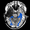
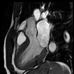
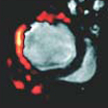
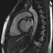
Within IST/e image acquisition focuses on the development and use of in vivo magnetic resonance imaging (MRI) and spectroscopy (MRS) techniques. These techniques offer exciting possibilities for the investigation of a range of structural, functional and metabolic parameters in living systems. Their non-invasive nature and versatility explain the important role of MR in biological and biomedical research. Not surprisingly, MR has gained an important position in human healthcare since its first arrival in medical centers in the late 1980’s.
The BioMedical NMR group headed by professor Klaas Nicolay and associate professors Gustav Strijkers and Jeanine Prompers has horizontal bore MR instruments for in vivo studies on live laboratory animals (field strengths of 7.0 and 9.4 Tesla), a 3.0 Tesla whole-body scanner for human MR research, and 0.47 and 1.41 Tesla MR instruments for contrast agent research. A well-equipped chemical laboratory is available for the preparation and characterization of MRI contrast agents. IST/e offers facilities for housing of and research on laboratory animals, for studies on cultures of mammalian cells, for biochemical and microscopic studies of excised tissues, and for advanced data processing, evaluation and visualization.